Your fish has worms – it’s a dreaded diagnosis for any aquarium enthusiast. Unfortunately,
worm infestations are common in fish, including beloved discus. These parasitic worms, known as helminths, can harm your fish by stealing nutrients, damaging tissues, and releasing harmful substances. This can lead to various illnesses, organ damage, and even death. Worms come in different types, including roundworms (nematodes), tapeworms (cestodes), and flatworms (trematodes). In this article, we will talk about the parasitic worm Capillaria pterophylli (Heinze, 1933) from the class Nematoda.
About Capillaria pterophylli
The worm is named after the fish Pterophyllum scalare, in whose intestines it was first discovered. It is an egg-laying, highly contagious (*) parasite capable of infecting not only angelfish but also any other fish in the aquarium, including discus fish (Symphysodon discus, Heckell, 1840).
Capillaria pterophylli is a small worm. Males reach 5-7 mm, females 9-16 mm in length. In my practice, I have encountered female worms in the intestines of discus fish reaching 18 mm. Despite their relatively large size, C. pterophylli is difficult to see with the naked eye, as they are very thin, like hairs, measuring only 0.04-0.09 mm in diameter.

Author
Katja Ekaterina Otstavnaia
Ichthyopathologist at eSHa

C. pterophylli parasites in the fish’s intestines, causing a disease known as capillariasis, manifested by inflammatory processes in the intestinal wall and digestive disturbances.
This leads to the fact that despite sufficient and varied nutrition, fish begin to lose weight, their activity decreases, the bright color of the body fades or darkens (depending on the discus breed), and in some cases, abdominal swelling is observed. Often, fish become extremely skittish, gather in a group, and stay
in the corner of the aquarium, getting scared when they see a person. Other symptoms may include “spitting out” food or refusing it, as well as the appearance of long semi-transparent feces with patches of mucous discharge. The appearance of long, semi-transparent feces with patches of mucous discharge
warrants special attention, as there is a common misconception about its cause among aquarium hobbyists.
For many aquarium enthusiasts, the presence of transparent, long feces in fish is considered a sign of a disease known as “hexamitosis.” However, this statement is incorrect and originated a few years ago, becoming the subject of careless copying by various internet resources. If you have observed this
symptom in your fish, it does not guarantee that they have developed hexamitosis. This symptom indicates a disruption in the functioning of your fish’s intestine, causing the mucous membrane of the intestinal wall to produce a large amount of mucus in response to an irritating factor. This irritating
factor can be the presence of unicellular flagellates from the group Flagellata, causing hexamitosis, as well as the presence of other unicellular or multicellular parasites, including worm infestations.
Therefore, if you have noticed such a symptom in your fish, you should not immediately resort to medications against hexamitosis. It is essential, first and foremost, to accurately diagnose the issue. This can only be reliably done using a microscope and conducting microscopic diagnostics of such long semi-transparent feces with mucous content. Based on my experience working with discus, angelfish, and other large cichlids, in 80% of cases, upon examining such feces, you will find eggs of the nematode Capillaria.
What do the eggs of Capillaria pterophylli look like?
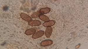
The eggs of C. pterophylli are clearly visible at 100x magnification under the microscope. They are elongated oval barrels of brown color with two lids on either side of the oval, measuring 0.05 mm in length. If you find even only one C. pterophylli egg in the examined sample, there should be no doubt that your fish are infected. Each day, mature female C. pterophylli produces dozens of eggs, which, along with feces, enter the aquarium water.
Development of Capillaria pterophylli
In 1952, Professor Heinz Hermann Reichenbach-Klinke determined that the development of C. pterophylli larvae after the eggs were released into the water with fish feces takes 33 days. Czech researcher František Moravec, in 1983, conducted experiments with Pterophyllum scalare angelfish, placing fish in an aquarium with C. pterophylli eggs. He found that to infect fish, the parasite does not require an intermediate host. In other words, infection occurs directly when the fish swallows the worm eggs. However, the researcher does not rule out that in the natural habitat, Oligochaeta worms may play a role as intermediate hosts for this parasite.
Another important discovery made by the scientist at that time, relevant for breeders and aquarium fish enthusiasts, was the establishment of the so-called prepatent period of development for this parasite, which is about 3 months at a water temperature of 20-23 degrees Celsius. That is, from the moment the fish swallows the egg until the larva hatches, grows, and develops into a mature adult, approximately 3 months pass at this temperature.
Parasite development in warmer water occurs faster, meaning the development period may be shorter in heat-tolerant discus fish. I conducted personal research with discus, angelfish, gouramis, and other fish species to determine the impact of parasites on them and treatment methods. Working with different groups of discus infected with C. pterophylli, I had to perform up to 4 treatments with a 2-week interval to rid the test fish of worms. As these are egg-laying worms, and their eggs can survive in the environment for some time, I additionally carried out preventive treatment every 3 months and examined their feces. After a year, I conducted a detailed study of test fish and found no worms in it.
Therefore, in the fight against capillariasis in discus and angelfish, I recommend a minimum of 4 treatments with a 2-week interval. Furthermore, if you have a microscope, monitor fish feces for the presence of eggs for 6 months after the last treatment (6 months because approximately in 3 months, the worm will become sexually mature and start laying eggs, and another 3 months later, the new generation of worms will become sexually mature and also start laying eggs).
(*) Contagiousness refers to the ability of a disease to be transmitted from one organism to another. This property can apply to bacteria, viruses, fungi, or other infectious agents. Contagiousness is determined by the ability of the pathogen to be transmitted through contact with an infected person, animal, or object.